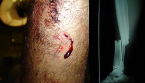
Open fractures are always assumed to be infected, and a small puncture wound is as liable as a laceration towards infection.
The most useful classification for open fracture is the Gustillo's classification :
a) Type I
It is essentially a low energy fracture.
There is only little soft tissue damage.
The wound is small (<1cm), and is clean
There is no comminution.
a) Osteosarcoma
Osteosarcoma, in it's classical intra-medullary type, is a highly malignant metaphyseal bone tumour which rapidly spreads to the periosteum and the surrounding soft tissues.
It is more common among children and adolescence, especially around the age of 15-25 years old.
Most commonly affects the long bones around the knee joint, for eg : distal end of femur, proximal end of tibia, and proximal end of humerus.
Introduction
Intervertebral disc comprises of 3 important components :
1) cartilage end-plate 2) nucleus pulposus 3) annular fibrosus
The cartilage end-plate is a thin articular plate situated in between the adjacent vertebral bodies and the disc proper. The disc receives nutrition from the vertebral bodies via these cartilage end-plates.
Nucleus pulposus is a gelatinous substance located slightly posterior to the central axis of the vertebra. It is surrounded by concentric fibro-cartilaginous rings, known as annular fibrosus.
In front and behind of the vertebral bodies and intervertebral disc are covered by strap ligaments, known as the anterior and posterior longitudinal ligament.
Introduction
Scaphoid fracture accounts for about 75% of carpal bone injury.
Common among young adults, rare in elderly and children.
The most common part of scaphoid bone fractured is the waist, which is the narrowest part.
Introduction
Patella is a sesmoid bone, with continuation from the quadriceps tendon, and patellar ligament.
It also receives insertion from vastus medialis and lateralis to it's medial and lateral edge.
The extensor snap is completed as the quadriceps muscles extends beyond the patella, and inserts into the proximal tibia (the medial and lateral retinacular fibres)
Introduction
Supracondylar fracture of the femur in young adults is a result of high energy injury, usually seen in road traffic accidents. Whilst in older age group, it may be an osteoporotic fracture.
Condylar fracture of the femur commonly associated with supracondylar fracture (fracture above the condyles extending into the knee joint), however, it can occur as an isolated fracture, for example fall from a height which results in the tibia thrusted into the intercondylar fossa of the femur.
Introduction
As in Rheumatoid arthritis, ankylosing spondylitis (AS) is a generalised chronic inflammatory disorder, which mainly involves the sacro-iliac joint (SI) and the spine. Typical presentation will be recurrent back pain and stiffness, with variable involvement of the shoulder, hip and other peripheral joints.
It's a male predominant disease (9:1), which typically affects teens and young adults aged between 15-25 years old. There is a strong familial aggregation and the genetic marker for AS is HLA-B27.
What you understand by union, non-union and consolidation?
Union is an incomplete repair, as the callus en-sheathing around the site of fracture undergoes calcification.
Palpation over the site usually causes slight tenderness, but attempts of moving the bone - the bone moves as one, and attempts of angulation causes pain.
X ray reveals that the fracture line is still visible, however, there is formation of fluffy bones (callus) around the periosteum that surround the fracture site.
Since repair is incomplete, stress towards the unprotected bone is avoided.
Monteggia-Fracture dislocation
Defined as fracture of the upper 1/3 of ulnar, together with dislocation of the head of radius.
The mechanism of injury is usually fall on an out-stretched hand with hyper-pronation of the forearm.
Occasionally, it can be caused by direct blow towards the back of forearm.
Clinical features
The deformity over the site of fracture (upper 1/3 of ulnar) is usually obvious.
However, the dislocated radial head can be obscured by the presence of swelling.
Pain and tenderness over the lateral aspect of elbow joint is suggestive.
It can be complicated as a radial nerve injury, causing wrist drop or injury to it's branch, the posterior interosseous nerve, causing finger drop.
First, examination should be started with inspection of the patient's back while he/she is standing.
a) Inspection - Posterior
1) Is there any shoulder asymmetry?
2) Is there any prominent crease over the flanks?
3) Any scoliosis?
Back pain is one of the commonest presenting complaint in an Orthopedic clinic.
Often, a definite diagnosis is not possible.
However, it is useful if we classify the problem under 3 headings :
- Sprengel’s – unilateral elevation of scapula; actually, failure of scapula to descend.
- Omovertebral bone – often seen with sprengel’s. Often associates Klippel-Feil syndrome, blocked vertebra, webbed neck
- Rhomboid fossa – a muscular impression on X-ray. No clinical consequence.
- Hemivertebra – only neatly triangular when drawn.
- Scrambled spine, also a structural scoliosis.
- Butterfly vertebra – missing primary ossification centers in the body, apparently migrating to the levels above and below.
- Intrathoracic rib – unlikely to produce any complications.
- Synostosis of rib (bifid rib, or costal synostosis). Failure to separate.
- Elongation of L5 TP, considered a physiologic variant most likely calcification of iliolumbar ligament.
- Spina bifida in the lumbar vertebra seems to associate with increased disc pathology.
- Reversed thoracic kyphosis or thoracic lordosis: recommendable to perform thoracic cage mensuration. If linked with a heart murmur, then straight back syndrome. If there is a heart murmur it is likely a result of floppy valve syndrome. High percentage have pathology requiring valve replacement.
- Costochondral ossification – physiologic calcification – everything (serum and tissue calcium) is normal.
- Thoracolumbar transitional ribs.
- Notochordal persistency resulting in cupid’s bow.
- Winking owl sign – metastasis (most common) or congenital. Sclerosis of the contralateral indicates congenital because it takes a long time to sclerose.
- There is an apparent correlation between spina bifida and DJD.
- Springboard divers, interior lineman, gymnasts are at high risk for acquired splondylolysis.
- If flexion/extension demonstrate 3% or more translation you confirm unstable listhesis; less than 3% does not rule in or out anything.
- Oppenheimer’s ossicles – nonunion of growth centers.
- Lordotic sacrum
- Injection granulomas [from arthritic injections in the gluteus] called dystrophic calcification (regularly seen in patients).
- Pseudotumor of the pelvis – results from observation of the growth plate between the ischial and pubic rami.
- Fabella – sesamoid-like bone in lateral gastroc often confused with synovial chondroplasia.
- Rockerbottom foot – missing tarsal bones
- Bipartate sesamoid bones.
- Tarsal coalition – bony bars in the tarsals.
- Pseudotumor of the humerus – lucency that results from the attachment of rotators – it disappears on baby-arm X-ray
- Supracondylar process – found on the humerus and points toward the elbow joint (normal variant). Often confused with osteochondroma – points away from the joint, found anywhere on the body, and has a chondral cap.
- Maudline’s deformity – ulnar deviation of wrist
- Ulna minus – short ulna. Non united growth centre of ulnar styloid process.
- Radius minus.
- Synostosis – failure of the bones to separate.
| Factor | Blastic Metastases | Paget’s Disease | Hodgkin’s Disease |
| Age (years) | >45 | >50 | 20-40 |
| Increased density | +++ | +++ | +++ |
| Expansion | ---- | +++ |
|
| Anterior Scalloping | ---- | ---- | +++ |
| Acid phosphatase | +++ | ---- | ---- |
| Alkaline phosphataes | ++ | +++ | ++ |
-
Pelvis includes protrusio acetabuli with Kohler’s teardrop and thickening of pelvic brim
-
Long bones: the tibia is the m/c lytic site
-
Complications
-
Deformity of bone include: Shepherd’ crook deformity of the proximal femur (coxa vara), Saber shin (ant. Tibial bowing), Protrusio acetabuli.
-
Cranial nerve neuropathies b/c foramen get smaller.
-
Linked to increased wear and tear, early DJD
-
Increased alkaline phospatase (up to 20x normal)
| Common Causes | Uncommon Causes |
| Osteoblastic metastasis | Sarcoidosis |
| Hodgkin’s Lymphoma | Chordoma |
| Paget’s disease | Myeloma |
| Degenerative sclerosis | Osteosarcoma |
| Osteomyelitis (fungal or chronic) | Ewings sarcoma |
| Idiopathic | Osteoid osteoma |
|
| Osteoblastoma |
|
| Bone island |
-Is a bone disease of unknown origin characterized by osteolysis followed by extensive attempts at repair.
-
Mimics: Paget’s disease mimics a Hemangioma. Hemangioma for the vertebral bodies produces a vertically striated pattern, which may closely mimic the vertical trabeculae of Paget’s disease.
-
Most likely site: Pelvis
Least likely site is the Fibula
-
List the 4 phases and describe each
-
Stage one: Osteolytic, Destructive, or Monophasic stage- Osteoclastic over activity creates gross loss of bone density described as osteoporosis circumscripta
-
Stage two: Combined, Mixed, or Biphasic stage – M/c encountered. Reflection of both destruction (Lytic) and production of bone (Blastic). Characterized by cortical thickening, increased radiopacity and accentuation of trabecular patterns with lucent areas mixed.
-
Stage three: Sclerotic or Ivory Stage – Uniform thickening of trabeculae with ivory appearance
-
Stage Four: Malignant Degeneration – Lethal stage
-
Clinical Features of Paget’s
-
2:1 males, m/c after 55
-
90% are asymptomatic. Pain, when present, is low intensity and may be associated with bowing deformities or fractures
-
Increased hat size because enlargement of the calvaria
-
Radiological features
-
Bone scan will be hot
-
Skull will demonstrate in earl lesions Osteoporosis circumscripta (described in c). More advanced or combined stage demonstrates cotton wool appearance, which is fuzzy, poorly defined edges of sclerotic areas.
-
Spine will demonstrate squared-off picture frame vertebra, which is thickened and enlarged vertebral endplates giving a squared look.
-
Homogenous increases radiopacity of vertebral body and creates an ivory vertebra
-
The 3 m/c causes of an ivory vertebra are: osteoblastic metastatic carcinoma, Paget’s disease, and Hodgkins lmphoma
-Can malignantly degenerate into chondrosarcoma
-
Key clinical features.
-
m/c primary benign tumor of the hand
-
Can be Solitary or multiple (called Ollier’s Disease)
-
Solitary enchondroma, is a benign tumor arising in the cartilage in the metaphysis as the physis goes away.
-
Usually a painless incidental finding
-
Most common complication is pathological fracture
-
Key radiological features.
-
The bone looks less dense do to cartilage tissue within the bone
-
40% will uptake Ca++, giving a speckled appearance- a round lesion with polka dots
-
What is Ollier’s disease? (Multiple enchondromatosis)
-
Enchondroma’s in multiple sites
-
Shares a common presenting complaint with what process?
-
Brodie’s Abscess
-
The radiolucent nidus of Brodie’s is much larger (>1 or 2cm)
-
Hole will be in the marrow
-
The halo rim of sclerosis surrounding the nidus is much thicker and more irregular
-
Key clinical features.
-
Pain at night relieved by aspirin
-
A “blister” on the bone between the cortex and the periosteum. Might show up as a hole or nidus, which is lucent.
-
Key radiologic features.
-
The nidus or hole is usually surrounded by sclerosis.
-A cortical bone tumor with a smooth contour and a continuous cortex
-Are found:
Ocuping parts of the skull that SHB spaces (sinuses)
Attached to the skull as a raised lesion
-
What syndrome includes the Osteoma as part of its classic presentation?
-
Gardener’s Syndrome
-Hemangioma, a primary benign neoplasm, is a slowly growing lesion of bone composed of newly formed capillary, cavernous or venous vessels.
-
Key clinical features
-
M/c primary benign tumor of the spine
-
No history of going malignant
-
Key radiologic features
-
Corduroy cloth or striated vertebra appearance which is accentuated vertical trabeculae.
-
Barrel shaped vertebra
-
In the skull,, it appears as one large lesion. Most occur in the frontal bone, creating a round or oval radiolucency, radiating sunburst or poked-wheel appearance
-lesion consisting of a cystic cavity filled with blood.
-
Key clinical features?
-
75% 5-20 yoa
-
Previous history of trauma. 80% occur in long bone (femur, tibia and spine)
-
Key radiological features
-
Only benign bone tumor known to cross the epiphyseal plate
-
Cortical ballooning or Blown-out appearance
-
Periostel buttressing at the edge of the lesion is characteristic.

 It is essentially a low energy fracture.