Possible aetiology of Acute pancreatitis :
Gall stones
Alcoholism
Post ERCP
Abdominal trauma
Complication of cardiothoracic, biliary and abdominal surgery
Hypercalcemia
Hyperparathyroidism
Pancreatic Divisum
Autoimmune pancreatitis
Scorpion bite
Drugs : Corticosteroids, Azathioprine, Thiazide diuretics
Mumps, Cocksakie viral infection
Idiopathic
History
Both males and females are equally affected.
Age of onset is around 4th - 5th decade of life.
History of any gall stone diseases, or alcoholism is important (2 most important cause)
Though rare, but ask for recent contact with children with mumps or cocksakie infection.
Symptoms are usually triggered after consumption of large meal or alcohol.
Patient usually complains of sudden onset of severe, continuous epigastric pain, which typically radiates to the back, relieved by bending forwards.
It's accompanied by excessive vomiting and retching, with persistent nausea in between.
Breathing and movements exacerbates the pain.
General examination
Patient appears ill, with shallow breathing.
If the patient looks pale, diaphoretic, it is likely that it has complicated as a hypovolemic shock.
There might be mildly tinged jaundice if the pancreatitis is caused by gall stones.
Even 2-3 days after the illness, the mild tinged jaundice can be caused by compression of biliary duct by edematous head of pancreas.
Shock features of tachycardia, hypotension.
Low grade fever may or may not present.
Abdominal examination
In acute pancreatitis, the complains of the patient may indicate severe pain, but there is usually minimal findings
during abdominal examination.
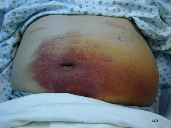
Cullen's sign
Grey Turner's sign
On inspection, if there's haemorrhagic pancreatits, there might be bruising (bluish purplish) discolouration over around the umbilicus (Cullen's sign) or left flank (Grey Turner's sign).
However, these signs are rarely seen nowadays.
The abdomen may not rise and fall with respiration, since the musculature is tightly contracted, and during onset of paralytic ileus.
Often there is accumulation of inflammatory exudates within the lesser sac, which eventually forms pseudocyst, suggested by epigastric fullness (distension), dullness during percussion over it.
Shifting dullness may be present, bowel sound may be reduced if there is pancreatic ascites.
Invesitgations
Acute pancreatitis is usually diagnosed by typical clinical presentation and laboratory investigation that reveals elevated serum amylase level. Serum amylase of 3-4 times greater than the normal level is suggestive of pancreatitis.
However, if serum lipase assay is available, it is more sensitive and specific.
Note that normal serum amylase level doesn't rule out Acute pancreatitis, and the level poorly correlates with the severity.
Both Ranson's and Glasgow's criteria is used to grade the severity of Acute pancreatitis.
If 3 or more factors are present in the patient, it indicates severe pancreatitis.
Imaging studies
Chest X ray, and plain abdominal X ray is not very helpful in the diagnosis of Acute pancreatitis
During early stages, if abdominal X ray is taken, non-specific signs such as Sentinel loop, Renal Halo sign or Colon cut off sign may be present.
Sentinel loop
Colon cut-off sign
Chest X ray may reveal pleural effusion, or if there is diffuse alveolar infiltrates, indicates ARDS.
Though Abdominal USG is non-diagnostic for Acute pancreatitis, but it must be done within 24 hours of presentation.
This is to rule out Acute cholecystitis as a differential diagnosis, to check whether the bile duct is dilated and to reveal any stones within the CBD (gall stone as a cause of Pancreatitis)
CT abdomen is not indicated in every patient.
Only when :
1) Diagnostic uncertainty
2) Severe pancreatitis
3) Clinical deterioration, with multi-organ failure, sepsis
4) Local complications occurs
Complications
Pancreatic pseudocyst
Management
a) Conservative
1) Gain IV access and rapid fluid resuscitation.
2) Give analgesics (usually IM pethidine is given)
3) Nil by mouth
4) Insertion of NG tube to relieve vomiting
5) Enteral feeding - nasojejunal tube (to maintain adequate nutrition)
6) Urinary catheterization is done
7) Monitor pulse, BP, urine output and CVP
8) Give antibiotics to prevent secondary infection
b) Endoscopic
If gall stone is strongly suspected as the cause of pancreatitis, the stones should be removed by basket (dormia) through endoscopic sphincterotomy.
If there is severe pancreatitis, or cholangitis occurs, both sphinterotomy and ERCP is done.
c) Surgery
Only indicated if : clinical deterioration during conservative management, unsure diagnosis, local complication occurs.
Adenocarcinoma of Pancreas
More common in males.
Age of presentation - 55-75 years old
85% of CA pancreas involves the head of pancreas, where the prognosis is usually poor.
Patients usually dies within 1 year of diagnosis (5 year survival is exceptional)
Upon presentation, most have progressed to a surgically incurable stage.
Though uncommon, but tumour arising from the distal CBD, duodenum and ampulla has a better outlook.
Currently, the risk factor of developing CA pancreas is thought to be :
Cigarette smoking
High fat and protein diet
Clinical features
Any elderly patient presents with painless jaundice, always suspect the possibility of Pancreatic cancer.
Typical symptoms are usually : Abdominal pain, jaundice and weight loss.
Jaundice is usually obstructive in nature, suggested by the classical triad of pruritus, clay-coloured stool and tea-coloured urine.
There might be steatorrhoea.
Abdominal pain - constant, dull aching, discomfort, over the epigastric region
Sought for symptoms of metastases
On examination, the gall bladder may be palpable, as accordance to the Curviosier's law, which states that,
"For patients presenting with clinically evident jaundice, and on examination the gall bladder is palpable, the cause is more likely to be due to Carcinoma of the Head of Pancreas".
Look for scleral icterus, hepatomegaly, etc.
Investigations
If stool occult blood test is positive, suggestive of a ampullary tumour.
Abdominal USG - usually done to look for liver metastases, or any mass lesion over the pancreas, and dilated bile ducts
CT abdomen - extent of metastases
To confirm the nature of obstruction, MRCP is preferred over ERCP since the former is less invasive.
Surgery of choice : Whipple's procedure


POST COMMENT
0 comments:
Post a Comment